Home
/ Animal Cell Mitosis Stages Microscope : Recognising The Stages Of Mitosis Aqa As Biology Revision Notes / • use a microscope to identify cells in interphase and different stages of mitosis from prepared animal and plant slides.
Animal Cell Mitosis Stages Microscope : Recognising The Stages Of Mitosis Aqa As Biology Revision Notes / • use a microscope to identify cells in interphase and different stages of mitosis from prepared animal and plant slides.
Animal Cell Mitosis Stages Microscope : Recognising The Stages Of Mitosis Aqa As Biology Revision Notes / • use a microscope to identify cells in interphase and different stages of mitosis from prepared animal and plant slides.. Animal mitosis plant mitosis have centrioles, centrosomes, and asters no centrioles. The end result of meiosis is four haploid daughter cells that each. Microscopes used in this lab do not have to be expensive or high powered. For example, within the nucleus lie the chromosomes. Dna supercoils and chromosomes condense (becoming visible under microscope).
Mitosis is part of the cell cycle where one cell divides into two identical daughter cells. The single strand of dna that makes up each chromosome produces an exact copy of itself. Animal cells are of various sizes and have irregular shapes. Nevertheless a number of mitotic stages can be defined: Prophase (b and 2), metaphase (c and 3), anaphase (mid 4 and late d and 5).
Mitosis Stages Of The Lily Microbehunter Microscopy from www.microbehunter.com Each chromosome is made of two genetically identical chromatids, joined by a centromere. Observing mitosis with fluorescence microscopy. The end result of meiosis is four haploid daughter cells that each. Mitosis, a phenomenon observed in all higher eukaryotes, is the mechanism that allows the nuclei of cells to split and provide each daughter cell with a figure 1 presents images from five of the mitosis stages, as well as the interphase period. Animal cells are of various sizes and have irregular shapes. This is the longest period of the complete cell cycle during which dna replicates, the centrioles divide, and proteins are actively produced. Mitosis is the process of cell division that forms two genetically identical nuclei from on parent cell nucleus. These undifferentiated cells undergo mitosis at a regular interval as the embryo increases in number of cells find and make observations of cells in each phase of mitosis in plant and animal tissue.
In animal cells, mitosis is characterized by the inward contraction of the cytoskeletal fibers and the pinching of the cell in a process called contractile cytokinesis.
In cell biology, mitosis (/maɪˈtoʊsɪs/) is a part of the cell cycle in which replicated chromosomes are separated into two new nuclei. The stages of mitosis is given below. You can prepare the slides of various stages of mitosis, including prophase, metaphase, anaphase and telophase. Mitosis animal cell under microscope stock photo edit now 1133754233 study of the stages of mitosis from permanent slides onion cell mitosis stages. This video takes you through microscope images of cells going through mitosis and identifies the different phases under the microscope and on a micrograph. This is the longest period of the complete cell cycle during which dna replicates, the centrioles divide, and proteins are actively produced. In the drawings below, you can see the chromosomes in the nucleus going through the process called mitosis, or division. These undifferentiated cells undergo mitosis at a regular interval as the embryo increases in number of cells find and make observations of cells in each phase of mitosis in plant and animal tissue. Both daughter cells will have the same number of chromosomes due to replication of dna stages of mitosis. I bought my twelve year old a microscope and these prepared slides for christmas, he has been looking for specimens to make. Each chromosome is made of two genetically identical chromatids, joined by a centromere. This inward contraction then produces a depression known as the cleavage furrow. Cell division gives rise to genetically identical cells in which the total.
Cell is enlarged in preparation for division. It is the stage in which the cell is growing in size and replicating its dna in preparation for division. Mitosis animal cell under microscope stock photo edit now 1133754233 study of the stages of mitosis from permanent slides onion cell mitosis stages. Repairing a damaged part of its body. Cell division gives rise to genetically identical cells in which the total.
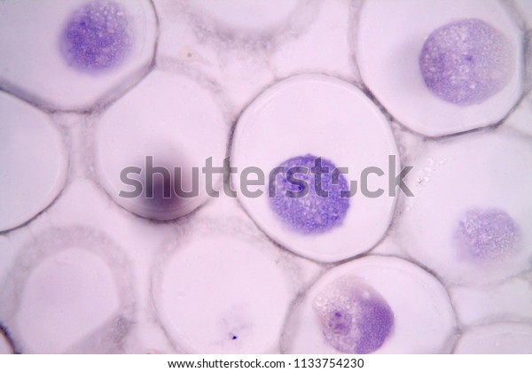
Mitosis Animal Cell Under Microscope Stock Photo Edit Now 1133754230 from image.shutterstock.com Mitosis is part of the cell cycle where one cell divides into two identical daughter cells. Root tip cells of onion. This video takes you through microscope images of cells going through mitosis and identifies the different phases under the microscope and on a micrograph. I bought my twelve year old a microscope and these prepared slides for christmas, he has been looking for specimens to make. Cell is enlarged in preparation for division. The single strand of dna that makes up each chromosome produces an exact copy of itself. Equipment used to photograph the onion root: Prophase (b and 2), metaphase (c and 3), anaphase (mid 4 and late d and 5).
Excellent value for your little scientists.
In the drawings below, you can see the chromosomes in the nucleus going through the process called mitosis, or division. This animation demonstrates the stages of mitosis in an animal cell. These undifferentiated cells undergo mitosis at a regular interval as the embryo increases in number of cells find and make observations of cells in each phase of mitosis in plant and animal tissue. This inward contraction then produces a depression known as the cleavage furrow. By examining the position of the chromosomes within the cell as well as looking for various other components of mitosis, you can discern the stage of mitosis you are viewing. In cell biology, mitosis (/maɪˈtoʊsɪs/) is a part of the cell cycle in which replicated chromosomes are separated into two new nuclei. Mitosis is the process of cell division that forms two genetically identical nuclei from on parent cell nucleus. In animal cells, mitosis is characterized by the inward contraction of the cytoskeletal fibers and the pinching of the cell in a process called contractile cytokinesis. Nevertheless a number of mitotic stages can be defined: Revise mitosis, the cell cycle and how stem cells work in humans and plants for gcse combined science, aqa. In this cell division, the two daughter cells have same number of chromosomes as that in the parent cells. Mitosis occurs in somatic cells of plants and animals. Most of the cells size range between 1 during mitosis the centrosome aids in dividing the cell and moving of the chromosome to the in addition the optical and electron microscope, scientists are able to use a number of other techniques.
In animal cells the centrioles separate and move apart, and radiating bundles of fibers, called asters, appear around them. Root tip cells of onion. • use a microscope to identify cells in interphase and different stages of mitosis from prepared animal and plant slides. • identify each stage of mitosis in the whitefish blastula or the onion root tip. The stages of mitosis is given below.
Leading The Change Uchim Uchitsya 9th Grade Lab Work Mitosis In Onion Root Tip Cells from 1.bp.blogspot.com Dna supercoils and chromosomes condense (becoming visible under microscope). Revise mitosis, the cell cycle and how stem cells work in humans and plants for gcse combined science, aqa. The process of mitosis consists of the following stages or phases For example, within the nucleus lie the chromosomes. Mitosis animal cell under microscope stock photo edit now 1133754233 study of the stages of mitosis from permanent slides onion cell mitosis stages. Both daughter cells will have the same number of chromosomes due to replication of dna stages of mitosis. I bought my twelve year old a microscope and these prepared slides for christmas, he has been looking for specimens to make. Mitosis, a phenomenon observed in all higher eukaryotes, is the mechanism that allows the nuclei of cells to split and provide each daughter cell with a figure 1 presents images from five of the mitosis stages, as well as the interphase period.
The cell is engaged in the metabolic activity and performing its prepare for mitosis (the next in animal cells, cytokinesis results when a fiber ring composed of a protein structure called actin around the center of.
In animal cells the centrioles separate and move apart, and radiating bundles of fibers, called asters, appear around them. Chromosomes are comprised of genetically identical sister chromatids (joined at a centromere). The single strand of dna that makes up each chromosome produces an exact copy of itself. • prepare your own temporary mounts of onion root tips and examine them under the microscope for different phases of the cell cycle. Microscopes used in this lab do not have to be expensive or high powered. This is an animal cell for sure. Repairing a damaged part of its body. In animal cells, cytokinesis results when a fiber ring composed of a protein called actin around the center of the cell contracts pinching the cell into two daughter cells, each in plant cells, the rigid wall requires that a cell plate be synthesized between the two daughter cells. In this cell division, the two daughter cells have same number of chromosomes as that in the parent cells. Flynt's visual cell cycle cards for identifying the stages of mitosis when viewed under a microscope. For example, within the nucleus lie the chromosomes. Dna supercoils and chromosomes condense (becoming visible under microscope). • identify each stage of mitosis in the whitefish blastula or the onion root tip.
Share :
Post a Comment
for "Animal Cell Mitosis Stages Microscope : Recognising The Stages Of Mitosis Aqa As Biology Revision Notes / • use a microscope to identify cells in interphase and different stages of mitosis from prepared animal and plant slides."


Post a Comment for "Animal Cell Mitosis Stages Microscope : Recognising The Stages Of Mitosis Aqa As Biology Revision Notes / • use a microscope to identify cells in interphase and different stages of mitosis from prepared animal and plant slides."