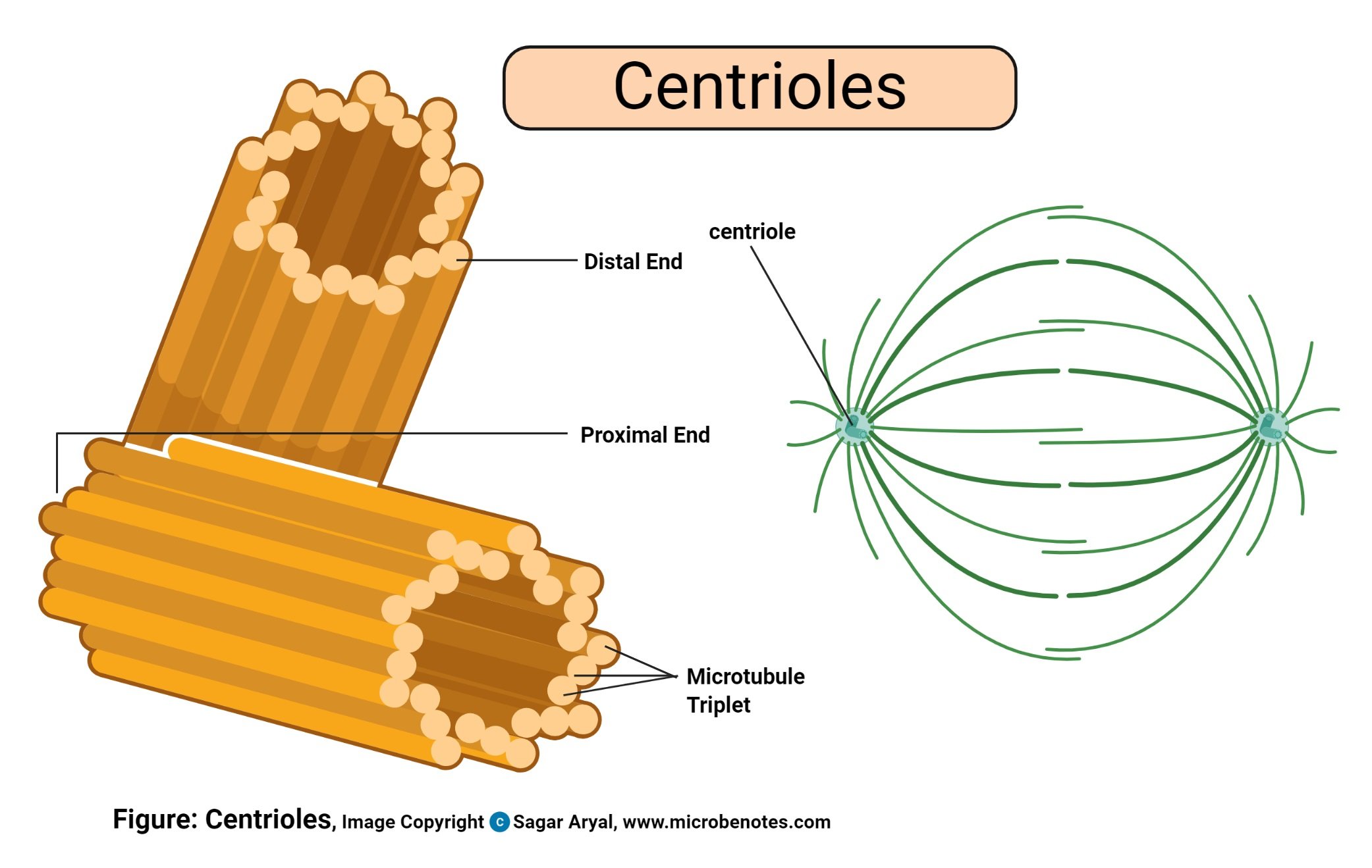
Animal Cell Diagram And Definitions : IB SEHS Topic 3 - Diagram of Mitochondrion and Generalized ... : Smooth endoplasmic reticulum, mitochondria, golgi bodies, lysosomes.. Each centriole is a ring of nine groups of fused microtubules. Cells are the starting point. In fact, most are invisible without using a microscope. There are three microtubules in each group. The plant cell as more rigid and stiff walls.
In truth, there are still features of plant and animal cells we're only lately. The parts of an animal cell have distinct functions. I spelt it wrong in the diagram, sorry). Plant cell and animal cell fall under eukaryotic type. The result is two centrosomes, each with its own pair of centrioles.
13 Best Images of Parts Of A Plant Cell Worksheet - Plant ... from www.worksheeto.com Cytoplasm, ribosomes, rough endoplasmic reticulum; Cells are the smallest units of life. This diagram shows a typical animal cell. The parts of an animal cell have distinct functions. Structure of generic animal cell all living organisms on earth are divided in pieces called cells. The result is two centrosomes, each with its own pair of centrioles. Animals, plants and microorganisms are always among us. While the plant cell resembles rectangular shape and.
Animals are made up of basic building blocks called the animal cell.
Table of contents definition explanation diagram structure types conclusion let us have a detailed overview of the animal cell, its types, diagram and structure. In truth, there are still features of plant and animal cells we're only lately. Printable animal cell diagram to help you learn the organelles in an animal cell in preparation for your test or quiz. The parts of an animal cell have distinct functions. During animal cell division, the centrioles replicate (make new copies) and the centrosome divides. These parts are called subcellular structures. This diagram shows a typical animal cell. I spelt it wrong in the diagram, sorry). The cell (from latin cella, meaning small room) is the basic structural, functional, and biological unit of all known organisms. Animals are made up of basic building blocks called the animal cell. Cells are the smallest units of life. But at the same time it is interpretive. Most cells are very small;
During animal cell division, the centrioles replicate (make new copies) and the centrosome divides. I spelt it wrong in the diagram, sorry). After completing this section, you should know: Cell is a tiny structure and functional unit of a living organism containing various parts known as organelles. It provides channels for the ribosomes to travel to.
Animal Cell- Definition, Structure, Parts, Functions and ... from microbenotes.com In truth, there are still features of plant and animal cells we're only lately. Most cells are very small; The animal cell diagram is widely asked in class 10 and 12 examinations and is beneficial to understand the structure and functions of an animal. Have a look at the micrograph of a nucleus and the diagram of the nucleus. Structure of generic animal cell all living organisms on earth are divided in pieces called cells. These parts are called subcellular structures. In fact, most are invisible without using a microscope. Just like us they are also living beings who perform various tasks similar to humans.
These parts are called subcellular structures.
Printable animal cell diagram to help you learn the organelles in an animal cell in preparation for your test or quiz. The largest organelle within the cell. Cell is a tiny structure and functional unit of a living organism containing various parts known as organelles. Let us look at animal cell parts and functions, using diagrams and illustrations. During animal cell division, the centrioles replicate (make new copies) and the centrosome divides. Since animal cells lack a rigid cell wall it allows them to develop a great diversity of cell types, tissues, and organs. This diagram shows a typical animal cell. It helps in carrying out the functions such as respiration, nutrition, digestion, excretion etc. An animal cell diagram is a great way to learn and understand the many functions of an animal cell. They are eukaryotic cells, that means they contain a membrane bound nucleus. In this video i'm going to draw labelled diagram of animal cell.in this video you will see the diagram of animal cell and it's labelling.this diagram of. Smooth endoplasmic reticulum, mitochondria, golgi bodies, lysosomes. Is the plumbing system of the cell but lacks ribosomes.smooth er plumb most of the cells waste and on a diagram can be is also another on of the plumbing system of the cell.
After completing this section, you should know: A system of flattened membranes called cisternae (mainpoint: The animal cell diagram is widely asked in class 10 and 12 examinations and is beneficial to understand the structure and functions of an animal. Lets us discuss the animal cell, types of an animal cell, animal cell diagram, its structure. The parts of an animal cell have distinct functions.
The Animal Cell parts - Biology 1000 with Ladapo at North ... from s3.amazonaws.com An animal cell diagram is a great way to learn and understand the many functions of an animal cell. The animal cell is more fluid or elastic or malleable in structure; In truth, there are still features of plant and animal cells we're only lately. Animal cell definition animal cells are the rudimentary unit of life for kingdom animalia organisms. With a light microscope you can see several structures inside the cell. This diagram shows a typical animal cell. Structure of generic animal cell all living organisms on earth are divided in pieces called cells. Unlike the eukaryotic cells of plants and fungi, animal cells do not have a cell wall.
During animal cell division, the centrioles replicate (make new copies) and the centrosome divides.
While the plant cell resembles rectangular shape and. In this video i'm going to draw labelled diagram of animal cell.in this video you will see the diagram of animal cell and it's labelling.this diagram of. The result is two centrosomes, each with its own pair of centrioles. All these work together to perform specific functions that are needed for. I spelt it wrong in the diagram, sorry). These structures are discussed in more detail in the following pages. Most cells are very small; Animal cell functions are solely dependent on the organelles and structures associated with the cell. These are organelles pertinent to plant cells. It helps in carrying out the functions such as respiration, nutrition, digestion, excretion etc. It provides channels for the ribosomes to travel to. Plant cells and animal cells fall under the eukaryotic category. Smooth endoplasmic reticulum, mitochondria, golgi bodies, lysosomes.
Share :
Post a Comment
for "Animal Cell Diagram And Definitions : IB SEHS Topic 3 - Diagram of Mitochondrion and Generalized ... : Smooth endoplasmic reticulum, mitochondria, golgi bodies, lysosomes."


Post a Comment for "Animal Cell Diagram And Definitions : IB SEHS Topic 3 - Diagram of Mitochondrion and Generalized ... : Smooth endoplasmic reticulum, mitochondria, golgi bodies, lysosomes."